Datei:Chlamydomonas TEM 09.jpg

Größe dieser Vorschau: 751 × 600 Pixel. Weitere Auflösungen: 301 × 240 Pixel | 601 × 480 Pixel | 961 × 768 Pixel | 1.280 × 1.023 Pixel | 1.800 × 1.438 Pixel.
Originaldatei (1.800 × 1.438 Pixel, Dateigröße: 784 KB, MIME-Typ: image/jpeg)
| Beschreibung |
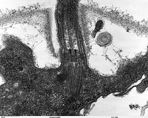
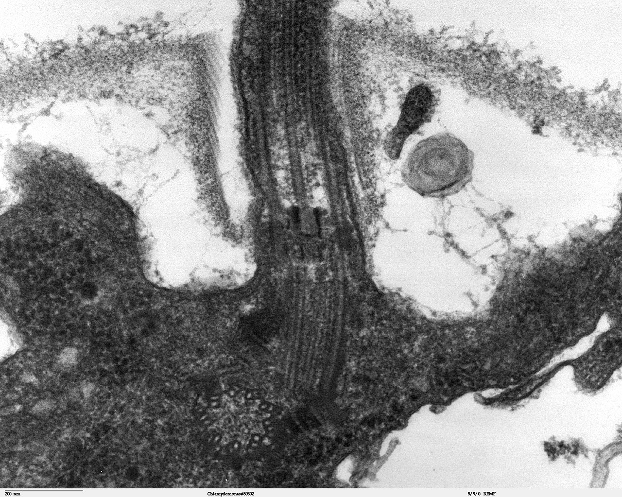
Transmission electron microscope image, showing an example of green algae (Chlorophyta). Chlamydomanas reinhardtii is a unicellular flagellate used as a model system in molecular genetics work and flagellar motility studies. This image is a longitudinal section through the flagella area. In the cell apex is the basal body that is the anchoring site for a flagella. Basal bodies originate from and have a substructure similar to that of centrioles, with nine peripheral microtubule triplets(see structure at bottom center of image). The two inner microtubules of each triplet in a basal body become the two outer doublets in the flagella. This image also shows the transition region, with its fibers of the stellate structure. The top of the image shows the flagella passing through the cell wall. |
| Datum | |
| Quelle | Source and public domain notice at http://remf.dartmouth.edu/imagesindex.html |
| Urheber | Dartmouth Electron Microscope Facility, Dartmouth College |
| Genehmigung (Weiternutzung dieser Datei) |
Released into the public domain |
| Dieses Werk wurde von seinem Urheber Dartmouth Electron Microscope Facility, Dartmouth College als gemeinfrei veröffentlicht. Dies gilt weltweit. In manchen Staaten könnte dies rechtlich nicht möglich sein. Sofern dies der Fall ist: Dartmouth Electron Microscope Facility, Dartmouth College gewährt jedem das bedingungslose Recht, dieses Werk für jedweden Zweck zu nutzen, es sei denn, Bedingungen sind gesetzlich erforderlich.
|
Dateiversionen
Klicke auf einen Zeitpunkt, um diese Version zu laden.
| Version vom | Vorschaubild | Maße | Benutzer | Kommentar | |
|---|---|---|---|---|---|
| aktuell | 06:47, 21. Sep. 2007 | | 1.800 × 1.438 (784 KB) | wikimediacommons>Neil916 | {{Information |Description= Transmission electron microscope image, showing an example of green algae (Chlorophyta). <br><br>''Chlamydomanas reinhardtii'' is a unicellular flagellate used as a model system in molecular genetics work and flagellar motilit |
Dateiverwendung
Die folgende Seite verwendet diese Datei: